

Visual fieldPerimetry
Maps the limits of your side vision. It is the core test for detecting glaucoma and following it over the years.
Ophthalmology investigations specialist
Ophthalmologist and Fellow of the International Council of Ophthalmology. My sub-specialty is the diagnostic side of eye care — OCT, visual field, Pentacam, biometry — running those scans, reading them, and reporting them.
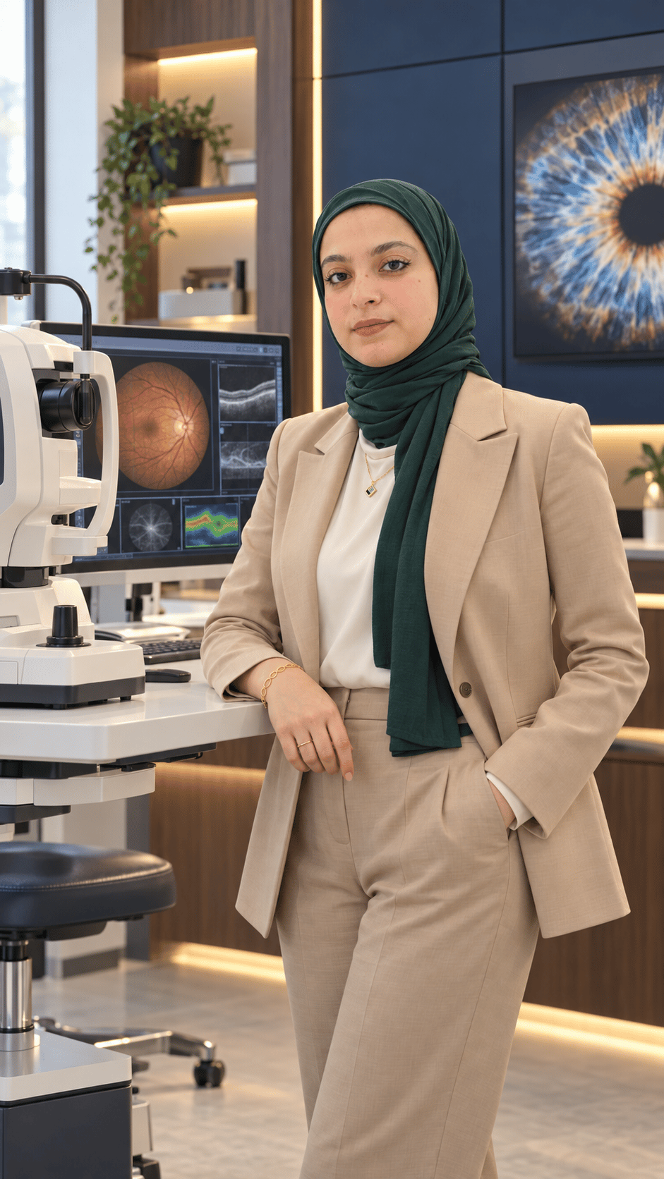
These scans show what an ordinary examination cannot reach, and they often find disease before you feel a single symptom. This is what I do every day.
The core scan
A light-based cross-section that shows the layers of your retina and optic nerve down to the micron — finer than any examination can see directly. It catches glaucoma early, before your sight is affected; it measures swelling at the centre of vision; and it tracks what diabetes is doing to the retina.
Nothing touches your eye. No injection, no pain. You sit at the machine for a few minutes and look at a target light, then we read the result together.
Maps the limits of your side vision. It is the core test for detecting glaucoma and following it over the years.
A full map of the cornea — its surface, thickness and curvature. Essential before laser vision correction, and for diagnosing and following keratoconus.
Precise measurement of the eye to choose the right lens power before cataract surgery.
Dye imaging of the retinal blood vessels, to locate leakage and blockage exactly before deciding treatment.
Sound imaging of the inside of the eye when a dense cataract or a bleed blocks the view to the retina.
Reading your scans and writing a clear medical report — whether the scan was done here or you arrive with a result from elsewhere.
For patients with diabetes
Diabetes can harm the retina for years before you notice anything. The early complications are silent — and they are exactly what an eye investigation is built to find.
Swelling at the centre of vision, diabetic macular edema, is often first found on a routine scan, while sight still feels normal. Found at that point, it can be treated and managed before it costs you vision.
The program brings together the examination and the scan a diabetic eye needs, in one visit.
Twice a year, even when your vision feels fine.
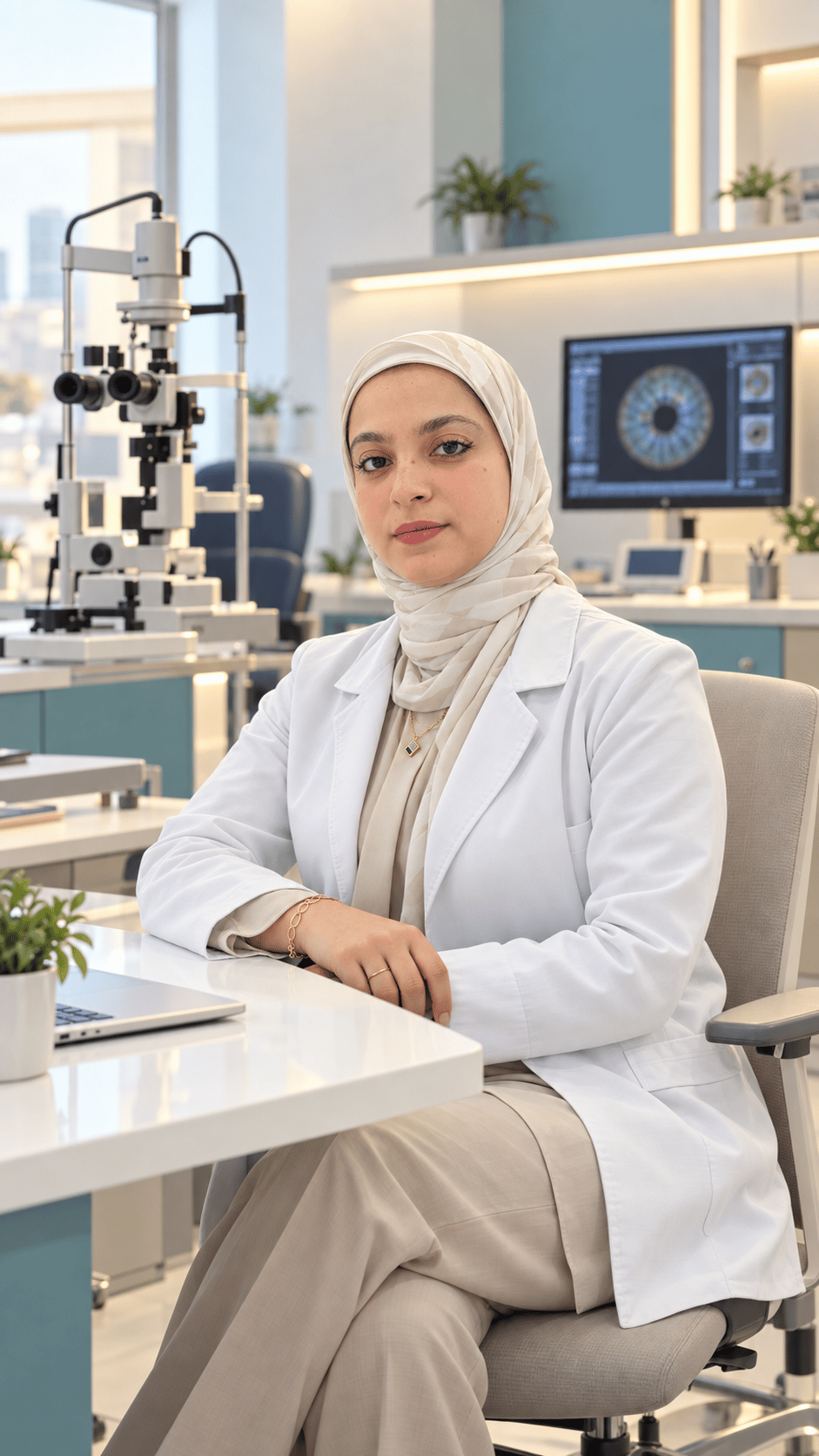
A full examination is where everything starts, whether you come with a specific complaint or for a routine check.
So you know what to expect, and how much time to set aside.
We talk through your complaint, your medication, and any general conditions such as diabetes or blood pressure.
The baseline measurements the rest of the examination is built on.
A close look at the front of the eye.
You may need drops to widen the pupil so I can see the whole retina. Vision then stays blurred and light-sensitive for four to six hours — arrange not to drive yourself that day.
Whatever scans your examination calls for.
We look at the images together, and you leave with a written report and a clear plan.
Dr. Reem graduated from the Faculty of Medicine at Ain Shams University in 2014, Very Good with Honours, and went on to a Master of Science in Ophthalmology at Al-Azhar University. Her thesis was on the corneal changes that come with diabetes.
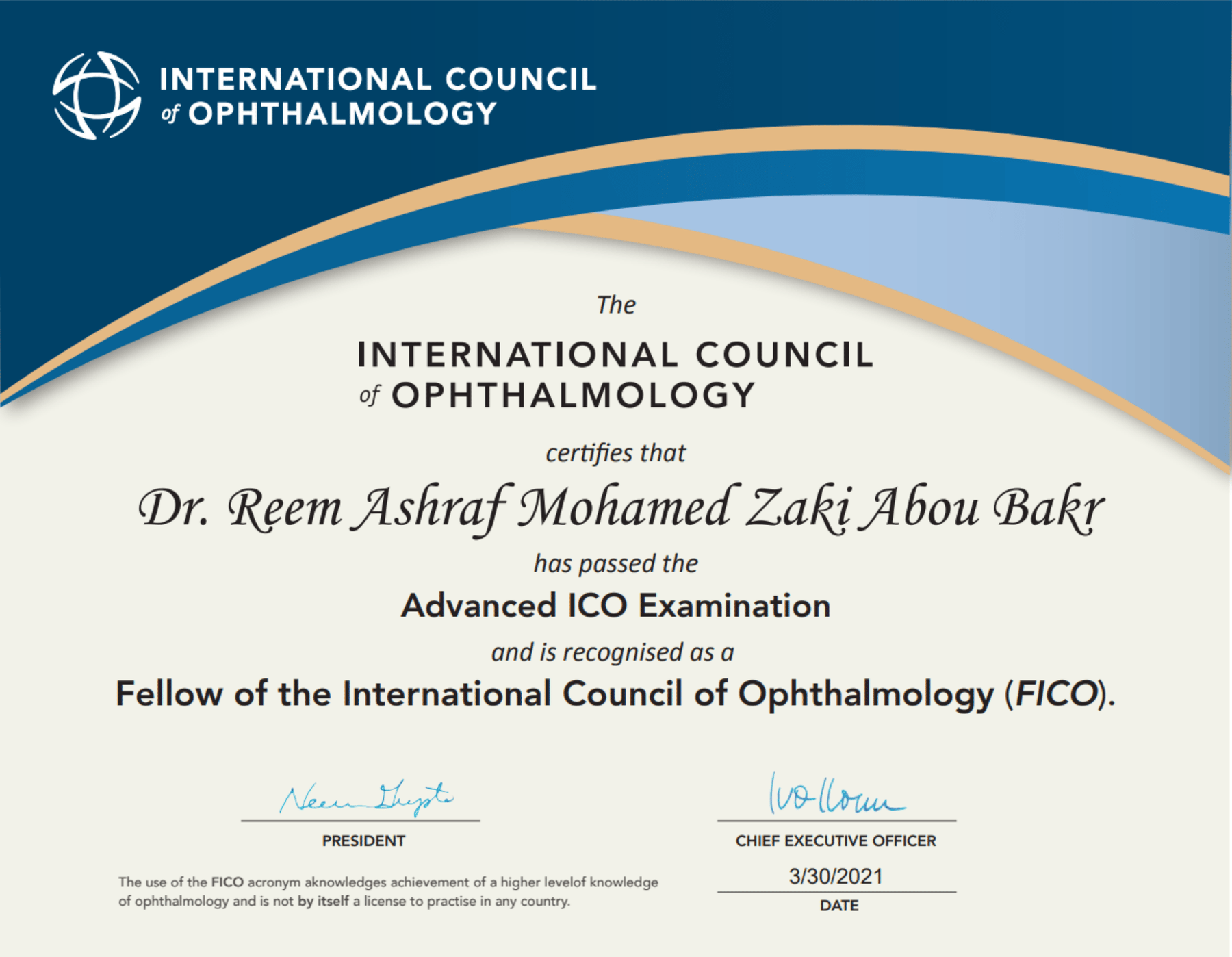
She then completed the International Council of Ophthalmology examinations — Basic Science and Optics, then Clinical Ophthalmology, then the Advanced Examination — and was awarded the Council Fellowship (FICO) in March 2021.
Since 2021 she has worked as Ophthalmology Investigations Specialist at Maadi Eye Specialized Center, after a year as an investigations fellow at Almashreq Specialized Hospital. Her days are spent running these scans, reading them and reporting them — and the diabetic eye has stayed her closest subject since that thesis.
Two locations, both by appointment.
Maadi
99 El Nasr St., Laselky Division, New Maadi
By appointment
New Cairo
Next to Hassan El Sharbatly Mosque, UMC Building — south of the Academy, New Cairo
By appointment
General answers to what patients ask most. They are here to explain, not to diagnose — your own eyes need an examination.
With no symptoms, a full examination every one to two years suits most adults, and a baseline examination at forty matters, because glaucoma and cataract become more common from then on. Children should have their vision checked before starting school. Diabetes, high blood pressure or a family history of glaucoma usually call for more frequent checks.
Yes. Diabetes damages the small blood vessels of the retina, and the early changes — including swelling at the centre of vision — cause no symptoms at all. By the time vision blurs, the damage is further along. Everyone with diabetes needs a retinal examination at least once a year; the Diabetic Eye Program runs every six months and adds an OCT scan.
Glaucoma is damage to the optic nerve, usually linked to the pressure inside the eye. The common form causes no pain and takes side vision first, so most people notice nothing until much is lost — and vision lost to glaucoma does not come back. Being over forty, a family history and high short-sightedness raise the risk. Eye pressure, an OCT of the optic nerve and a visual field test find it early, when treatment can slow or stop it.
It is safe and painless. OCT uses light, not radiation, and nothing touches the eye. You look at a target for a few minutes while the machine scans, and it can be repeated as often as follow-up needs.
In keratoconus the cornea thins and bulges into a cone, blurring and distorting vision. It usually begins in the teens or twenties, and eye rubbing and allergy are linked to it; a prescription that keeps changing, especially rising astigmatism, is a warning sign. Laser correction on a keratoconic cornea can make it worse, so a Pentacam map is taken first to rule it out. Found early, treatment can stop it progressing.
A cataract is clouding of the eye’s natural lens. It comes mostly with age, though diabetes, steroid medicines and injury bring it on earlier. Vision turns hazy, colours fade and glare at night gets worse. When it interferes with daily life, surgery replaces the cloudy lens with a clear artificial one — and biometry measures the eye beforehand to choose that lens’s power, which decides how well you see afterwards.
See an ophthalmologist the same day for sudden loss or dimming of vision, even if it recovers; a sudden shower of floaters or flashes of light; a shadow or curtain across part of your vision; a painful red eye, especially with blurred vision, headache or nausea; or any injury to the eye. After a chemical splash, rinse the eye with plenty of clean water first, then come in.
An undilated pupil shows only a small part of the retina. The drops widen it so the whole retina, out to its edges, can be examined — essential in diabetes, after flashes or floaters, and in short-sighted eyes. Vision stays blurred and light-sensitive for four to six hours, so bring sunglasses and arrange not to drive yourself.
Screens do not damage the eye, but long sessions reduce blinking, which leaves the eyes dry, tired and strained. Every twenty minutes, look at something about six metres away for twenty seconds. In children, more time outdoors is linked to slower progression of short-sightedness.